Highlights
┬Ę The prevalence of patients with central precocious puberty (CPP) has rapidly increased.
┬Ę Significant associations were observed between abnormal pituitary findings on MRI and CPP patients.
┬Ę Careful consideration of MRI screening is needed in patients with CPP.
Introduction
Central precocious puberty (CPP) is defined as abnormal onset of puberty before 8 years of age in girls and before 9 years of age in boys due to early activation of the hypothalamic-pituitary-gonadal (HPG) axis [1]. Approximately 85%ŌĆō90% of girls with CPP are idiopathic in nature, while up to 33% of girls with CPP and 40%ŌĆō90% of boys with CPP have pathological brain lesions [2-4]. According to a consensus statement on precocious puberty, all boys and girls aged <6 years with CPP should undergo brain magnetic resonance imaging (MRI) [5]. Moreover, brain MRI is required in girls with neurological symptoms and rapid pubertal development because they are more likely to have pathological intracranial lesions [5]. However, whether all girls who develop CPP between the ages of 6ŌĆō8 years require brain MRIs remains controversial. Due to the low likelihood of pathologic lesions, most opinions are against performing MRI in girls diagnosed with CPP after 6 years of age and in those without neurological symptoms [6-8]. Nevertheless, the importance of routine brain MRI in girls with CPP has been emphasized because of the likelihood of tumor or other incidental findings that cannot otherwise be predicted [6,7,9].
In 2018, the Korean Ministry of Health and Welfare expanded health insurance coverage for brain MRI in patients with CPP to help identify pathological brain lesions. Therefore, this study aimed to investigate the prevalence of MRI abnormalities and to evaluate the need for routine brain MRI in patients newly diagnosed with CPP. In addition, we examined the associations between the prevalence of pituitary abnormalities and clinical and laboratory factors in patients with CPP.
Materials and methods
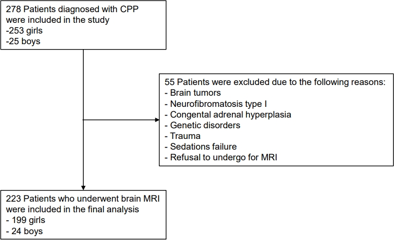
We retrospectively reviewed the medical records of patients diagnosed with CPP between March 2020 and September 2021. Among the 278 patients with CPP, 223 who underwent brain MRI were included in this study. CPP was diagnosed by early onset of secondary sexual characteristics before the age of 8 years for girls and 9 years for boys with peak luteinizing hormone (LH) level Ōēź5.0 IU/L by gonadotropin-releasing hormone stimulation test. Nonetheless, based on the age at the time of diagnosis, we included CPP patients, girls under the age of 9 years, and boys under the age of 10 years.
We recommend brain MRI for all patients diagnosed with CPP. Patients with previously known brain tumors, neurofibromatosis type I, congenital adrenal hyperplasia, genetic disorders, trauma, sedation failure, or parent refusal to allow them to undergo brain MRI were excluded from the study (Fig. 1).
Patient height and weight were measured with a Harpenden stadiometer (Holtain Ltd., Crymych, Wales, UK) and a digital scale with precisions of 0.1 cm and 0.1 kg, respectively. Weight and height standard deviation scores (SDSs) were calculated based on the 2017 Korean National Growth Charts, while body mass index (BMI) was calculated using a standard formula (kg/m2) [8]. Bone age (BA) was assessed using the Greulich-Pyle method. In addition, plasma estradiol (E2) and basal and peak serum LH and follicle-stimulating hormone (FSH) levels were measured using an immunoradiometric kit (Beckman Coulter, Brea, CA, USA).
To determine the significance of incidental MRI findings in patients with CPP, 90 age-matched girls who visited the pediatric neurology clinic for headache and underwent brain MRI were enrolled as controls.
High-resolution MRI was performed before and after gadolinium-enhanced T1-weighted imaging with coronal and sagittal sections. Dynamic contrast MR images using thin sections (2 mm) were acquired for the sellar region. In addition, T2-weighted images of the entire brain were acquired for nonpituitary incidental findings.
Positive MRI findings were classified into 3 categories: (1) abnormal pituitary findings (brain lesions located close to the pituitary gland); (2) nonpituitary incidental findings (lesions that are not closely located to or involve the pituitary gland); and (3) pathological findings (those related to CPP or headache; causative brain lesions that may cause headaches).
1. Statistical analysis
The results of descriptive statistics were reported as mean┬▒ standard deviation after the test of data normality. The chisquare test and Fisher exact test were used to compare the numbers and prevalence of MRI findings between the groups. Unadjusted odds ratios for MRI abnormalities between the CPP and headache control groups were calculated using the chi-square test. Multivariate regression analysis was performed to determine the statistical significance of the biochemical and laboratory factors in the brain MRI findings of patients with CPP. IBM SPSS Statistics ver. 21.0 (IBM Co., Armonk, NY, USA) was used to perform the statistical analyses. Significance was identified as values of P<0.05.
Results
1. Clinical characteristics and brain MRI findings in CPP cases
Among the 223 eligible patients with CPP who underwent brain MRI screening, 199 (89.2%) were girls (mean age, 8.22┬▒0.05 years) and 24 (10.8%) were boys (mean age, 9.41┬▒0.64 years) (Table 1). The patientsŌĆÖ baseline characteristics, including mean age at diagnosis, Tanner stage, difference between BA and chronological age (CA) (BAŌłÆCA), basal and peak LH and FSH levels, and peak LH/FSH ratio are listed in Table 1.
Among all patients with CPP, 84 (37.7%) exhibited positive MRI findings, while 139 (62.3%) had normal MRI findings. Pituitary findings were abnormal in 54 patients (24.2%), in whom the prevalence of Rathke cleft cyst (RCC) was highest (16.1%). Incidental nonpituitary findings were observed in 29 patients (13.0%); among them, pineal cysts were the most common (9.0%). Furthermore, only 1 girl (6.1 years of age) had a pathological brain lesion, which was diagnosed as a hypothalamic hamartoma (Table 2).
2. Comparison of brain MRI findings of patients with CPP by sex and age at diagnosis
No significant differences were observed in the prevalence of pituitary abnormalities or nonpituitary incidental findings between boys and girls with CPP (P=0.361 and P=0.938, respectively) (Table 2).
Among the girls enrolled in the study, 55 (27.6%) were aged 6 to < 8 years and 144 (72.4%) were aged 8 to <9 years (Table 3). Comparison of data by age among girls with CPP revealed no significant differences in the prevalence of pituitary abnormalities at the time of CPP diagnosis (P=0.947). Similarly, no significant difference in the prevalence of nonpituitary incidental findings was identified based on age at diagnosis in girls with CPP (P=0.577) (Table 3).
3. Abnormal brain MRI findings in girls of the CPP group versus the headache control group
The mean age of the 90 girls in the headache control group was 8.5┬▒0.12 years. MRI findings were normal in 74.4% and positive in 25.6% of girls in the headache control group. Approximately 7.8% of girls in the headache control group exhibited abnormal pituitary findings on the brain MRI, whereas 25.1% of the girls in the CPP group had abnormal pituitary findings (Table 4). Significant associations were observed between abnormal pituitary findings on MRI and CPP, with OR 3.979 (95% confidence interval, 1.726ŌĆō9.173), not adjusted for age or Tanner stage. Among the abnormal pituitary findings, RCC was most significantly associated with CPP, with unadjusted OR 5.976 (P=0.001).
4. Correlation between clinical and biochemical characteristics and MRI findings
No significant differences in laboratory findings, including basal LH, basal FSH, peak LH, and peak FSH level or peak LH/FSH ratio, were identified between patients with CPP and abnormal versus normal MRI findings. None of the clinical characteristics or auxological data, including height SDS, weight SDS, BMI SDS, puberty stage, BA SDS, or BAŌłÆCA, were significantly correlated with pituitary abnormalities on MRI (data not shown).
Discussion
It has been reported that OW children experience earlier In this study, a relatively higher number of positive brain MRI findings was observed in patients with CPP. Among pituitary abnormalities, RCC exhibited the highest prevalence. However, pathological intracranial lesions related to CPP were rare (0.4%).
In a 2003 European study, occult intracranial lesions were identified in 8% of CPP patients. However, its prevalence decreased with age and was 26.85% in patients aged <6 years, 2.17% in those aged 6ŌĆō6.9 years, and 1.66% in those aged 7ŌĆō7.9 years [10]. A study of 208 girls with CPP in Copenhagen performed from 1993ŌĆō2009 showed incidental findings, including pineal cysts and pituitary microadenomas, in 20 (9.6%) girls [6]. Our study demonstrated a higher percentage of positive MRI findings in patients with CPP compared to previous studies. Furthermore, the prevalence of pituitary abnormalities was higher in girls with CPP than in those in the control group (25.1% vs. 7.8%). Moreover, our study showed no significant age-related differences in the frequency of positive MRI findings. No significant sex-based differences were reported in the prevalence of pituitary abnormalities or nonpituitary incidental findings. This is probably due to the small sample size, especially that of boys, and that none of the girls enrolled in our study were younger than 6 years.
Previous studies reported a possible association between CPP and RCC. Jung et al. [11] reported that 36 of 91 patients with CPP had RCC (39.5%) on brain MRI. Lim et al. [12] reported that 18% of patients with symptomatic RCC had CPP. In our study, 20% of patients with CPP had RCC, and a female predominance was noted. Additionally, among the pituitary abnormalities, RCC was significantly associated with CPP. Additionally, compared to headache controls, RCC was most significantly associated with CPP among pituitary abnormalities.
Upon comparing the MRI findings of girls with CPP to those with headache, Kim et al. [13] reported a higher prevalence of incidental hypothalamic-pituitary lesions in girls with CPP. However, although RCC was the most frequent incidental hypothalamic-pituitary lesion, the authors could not establish a relationship between RCC and CPP.
RCC is a benign nonneoplastic cyst that originates from Rathke's pouch's remnants. However, its clinical relevance and proper management in pediatric patients are not clear. Although RCC was noted in up to 12%ŌĆō33% of autopsies, it is asymptomatic in most cases [12,14]. The most common clinical manifestation is headaches, while other manifestations include visual field defects and pituitary endocrine dysfunction [14,15]. None of the patients enrolled in this study had any symptoms other than CPP.
Several studies have investigated the relationship between CPP and RCC. However, the exact mechanism of precocious puberty remains unknown [16-18]. In addition, more studies are needed to determine the relationship between RCC size and CPP degree. In our study, the follow-up MRI performed for 3 patients with RCC revealed no increase in lesion size over time.
Pineal cysts were the most frequent nonpituitary incidental MRI finding in our study. Previous studies demonstrated pineal cysts in 1.1%ŌĆō4.3% of brain MRI examinations and reported a female predominance that caused non-specific symptoms [19,20]. Although pineal cysts exhibited a higher prevalence in patients with CPP compared to headache controls, none of the CPP patients with pineal cysts showed neurological symptoms at diagnosis. Recent studies have revealed that the underlying growth tendencies of pineal cysts are very low [21], and that we should consider pineal cysts as incidental lesions even though they were observed more commonly in the CPP group.
None of the patients with CPP diagnosed with microadenoma or arachnoid cysts exhibited neurologic or endocrine symptoms other than CPP. Although there are limited data on long-term outcomes, none of the CPP patients received surgery or showed other hormonal abnormalities due to brain lesions such as microadenomas, arachnoid cysts, and partial empty sellas as incidental findings.
Compared to other recent studies, our data showed relatively higher positive MRI findings in CPP patients compared to the headache controls [13]. However, all studies including ours had a low prevalence of pathologic lesions in CPP. Furthermore, information such as BA and hormone levels were not obtained from the headache control group, limiting establishment of the exact relationship between positive MRI findings and CPP.
The prevalence of pathological MRI findings was lower in our study compared to previous studies. Mogensen et al. reviewed the MRI findings of 229 girls with CPP; of them, 6.3% without neurological signs or symptoms had pathological lesions on MRI [6]. Pedicelli et al. identified pathological lesions in 3% of patients with CPP versus incidental findings in 11% of patients [9]. However, recent studies reported a lower prevalence of pathological lesions on MRI. A pathological prevalence of 0.4% was observed in a Taiwanese study of 403 girls with CPP, questioning the need for routine brain MRI in all girls diagnosed with CPP, especially for those who do not exhibit neurological symptoms [22]. Similar to the prevalence of pathologic brain findings, our data revealed a decreasing trend in the prevalence of pathological findings on brain MRI. The incidence of CPP has steadily increased in recent years, possibly because of increased awareness of the condition and earlier activation of the HPG axis [23]. This may explain the lower prevalence of pathological neuroimaging findings in patients with CPP in recent studies, including ours.
Previous studies evaluated the associations between clinical and biochemical factors and MRI findings in patients with CPP and reported findings similar to ours. Mogensen et al. reported that clinical and biochemical factors were insufficiently sensitive or specific for predicting brain abnormalities in girls diagnosed with CPP [6]. Similar to a recently published retrospective study of 295 girls in Korea, our study data revealed that the clinical characteristics and biochemical parameters of our cohort were not significantly associated with positive MRI findings [13].
This study had some limitations. First, the mean age of the enrolled patients was higher than previous studies. A few older patients may have normal variants of early puberty rather than true CPP. In addition, none of the CPP girls enrolled in this study were diagnosed before 6 years of age. Second, the sample size was relatively small, especially that of boys, owing to the short study duration. This may explain why no significant sexbased differences in positive MRI findings were observed in our study. Furthermore, CPP boys were excluded from statistical analysis between the CPP and headache control groups, and our study does not reflect all CPP patients enrolled in the group. Finally, different brain MRI protocols of the headache control group with CPP patients may be another limitation. However, the brain MRI protocol at our institution is sufficient for detecting Rathke cleft cysts and partial empty sellas. In addition, patients suspicious for microadenoma on initial brain MRI by the pediatric radiologist underwent additional dynamic imaging for pituitary fossa pathology.
In conclusion, true pathological findings are rare on brain MRI in patients with CPP. Considering its high cost, routine brain MRI screenings in patients with CPP should be carefully considered after explaining its risks and benefits.